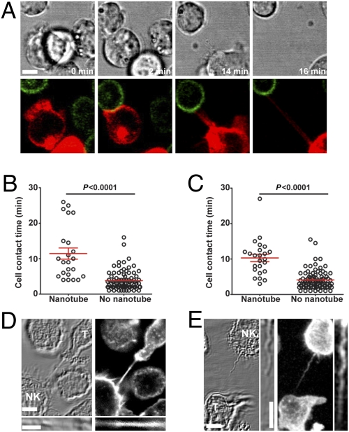
Fig. 2.
Characteristics of human NK cell membrane nanotubes. (A) Time-lapse microscopy reveals the formation of a nanotube between NKL labeled with membrane dye DiD (red) and P815/MICA-YFP (green, n > 100). Images acquired by time-lapse microscopy of primary NK cells (n = 109) (B) and NKL cells (n = 110) (C) coincubated with P815/MICA-YFP were analyzed to record the length of time of contact between cells and then whether or not membrane nanotubes formed as cells departed. Cocultures of NKL (labeled NK) and P815/MICA-YFP (labeled T for target cell) were fixed and stained with phalloidin-Alexa633, which marks f-actin (white, n > 100) (D) or anti-α-tubulin (E), followed by secondary mAb conjugated to Alexa633 (white, n = 78). Thin panels show an enlarged view of the nanotube. (Scale bars: 10 μm.)

