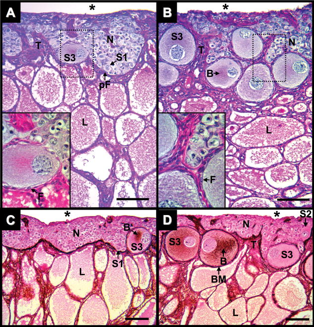
Fig. 3.
Ovary of Alligator mississippiensis in parasaggital view. A: Three months after hatching stained with PAS/AB. B: Five months after hatching stained with PAS/AB. C: Three weeks after hatching stained with PAMS. D: Five months after hatching stained with PAMS. Dotted line rectangles define areas shown in greater magnification in lower left of images. Scale bars = 100 um. Nest of germ cells (N); connective tissue trabeculae (T); coelomic cavity (*); stage-1, 2, and 3 oocytes (S1, S2, and S3, respectively); follicular cells (F); prefollicular cell (pF); lacunae (L); Balbiani bodies (B); and basement membrane (BM).

