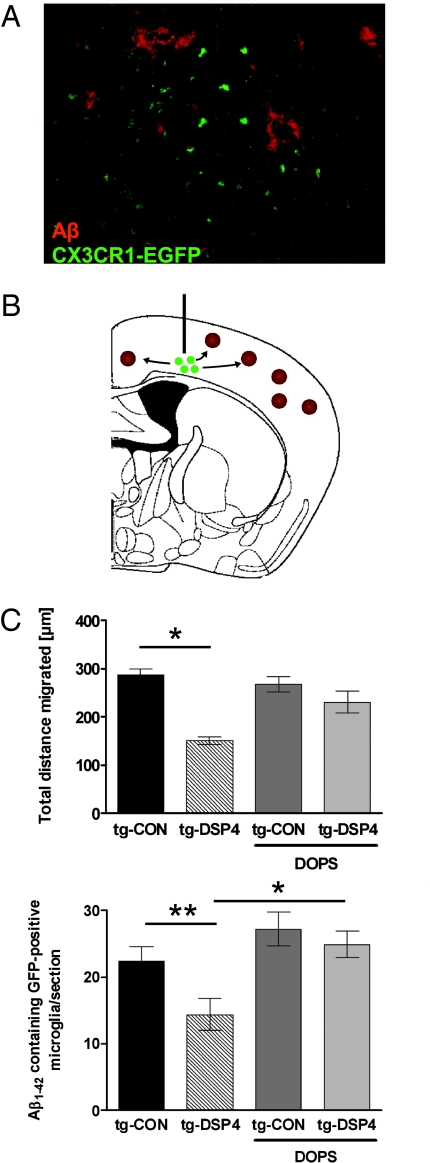
Fig. 4.
Decrease of microglial migration after NE depletion in vivo is rescued by the NE-precursor L-threo-DOPS. (A) APPV717I-transgenic mice treated with DSP4 or solvent control received a single injection of primary murine microglia derived from CX3CR1-EGFP-transgenic mice. A subgroup of animals in both DSP4-treated and control groups received three i.p. injections of the NE precursor L-threo-DOPS (DOPS) over 24 h to increase NE levels within the neocortex. Shown is confocal laser scanning microscopy of Aβ plaques and EGFP-positive microglial cells. (Scale bar: 50 μM.) (B) Scheme of the intracerebral injection site depicting the migration of EGFP-positive microglial cells (green) toward amyloid plaques (brown). (C) The total distance migrated and the number of EGFP-positive microglial cells per section was evaluated by analyzing serial sections with a defined distance to each other and the injection site (n = 6 ± SE; *, P < 0.05; **, P < 0.01, one-way ANOVA, Tukey's post hoc test).

