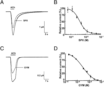
Fig. 2.
Inhibition of ACh-evoked currents from neuronal and muscle-type nAChRs by the phycotoxins. SPX with human α4β2 expressed in oocytes (A and B); GYM with Torpedo α12βγδ incorporated into the oocyte membrane (C and D). (A and C) Typical inward nicotinic currents evoked by a 5-s perfusion of 150 μM ACh (EC50 value for α4β2) before and after application of 1.5 nM SPX (A), and a 7-s perfusion of 25 μM ACh (EC50 value for α12βγδ) before and after application of 1.5 nM GYM (C). The desensitization component typical of α12βγδ is not modified in presence of the toxin. (B and D) SPX (B) and GYM (D) concentration-to-current inhibition relationships. The amplitudes of the ACh-induced currents recorded in the presence of SPX and GYM (mean ± SEM; 3–4 oocytes per concentration) were normalized to control currents and fitted to the Hill equation. SPX on α4β2: IC50 = 3.87 ± 1.1 nM; nH = 1.9 ± 0.33; GYM on α12βγδ: IC50 = 2.8 ± 1.15 nM; nH = 0.96 ± 0.15 (compare Table 2).

