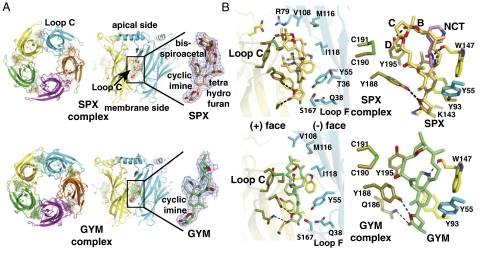
Fig. 3.
Overall views of the A-AChBP pentameric complexes and subunit interfaces with bound phycotoxins. (A) Ribbon diagrams of the pentamer (Left) and subunit interface (Center) with bound SPX (Upper; orange bonds and surface, red oxygens, blue nitrogen) and GYM (Lower; green bonds and surface, red oxygens, blue nitrogen) viewed from the “membrane” side (Left) and radial perspectives with the apical side at top and the membrane side at bottom (Center). The main and side chains at the (+) and (-) faces of the interface are displayed in yellow and cyan, respectively. The bound toxins (Right) are perfectly ordered, as shown by their 2.5/2.4 Å resolution 2Fo-Fc electron density maps contoured at 1.2σ (blue). (B) Close-up views of the bound toxins in their aromatic nest at the subunit interface, showing details of the cyclic imine environment. Bound SPX (Upper) is shown alone (Left) and overlaid with nicotine as bound to L-AChBP (pink bonds; Right). Bound GYM (Lower) is shown alone. The dashed lines denote H bonds.

