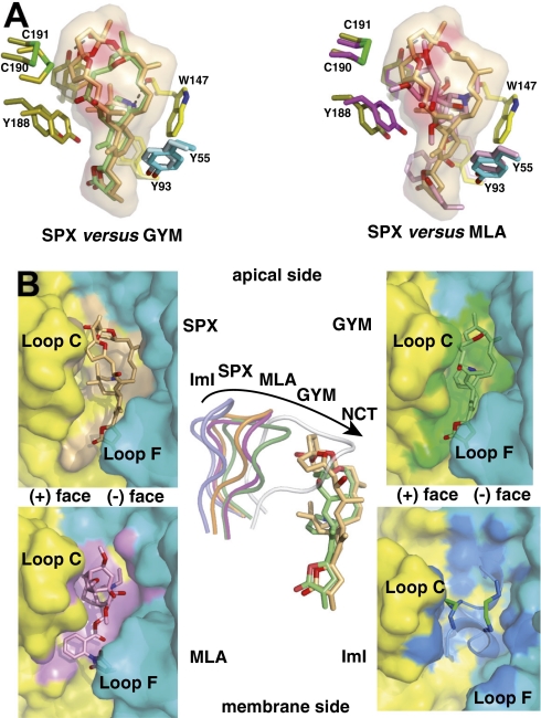
Fig. 4.
Structural comparisons of the SPX and GYM complexes with other antagonist complexes. (A) Superimposition of SPX (orange toxin) and GYM (green) (Left) and SPX (orange) and MLA (pink) (Right) bound to A-AChBP. The molecular surface of SPX and key side chains within the binding pocket are displayed. (B Left and Right) Molecular surfaces buried at the A-AChBP subunit interfaces by bound SPX (orange toxin and surface), GYM (green toxin and surface), MLA (pink toxin and surface), and the peptidic α-conotoxin ImI (blue toxin with green disulfides, mauve surface), viewed radially from the pentamer outer periphery. (B Center) Overlay of loop C in the SPX (orange loop), GYM (green), MLA (magenta), ImI (blue) and nicotine (white) complexes as viewed from the pentamer apical side, also showing overlaid bound SPX and GYM molecules.

