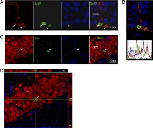
Fig. 3.
Phenotypic characterization of the BrdU-positive cells in mouse dentate gyrus. (A) An illustrative BrdU+ cell within the dentate gyrus (DG) is labeled with DCX and NeuN (arrowheads), indicating transition from a young to mature neuron and migration toward the granular cell layer (GCL) 24 h after APα and BrdU injections. (B) Confocal laser scanning histogram of IR fluorescent intensity profile verification of colocalization. DCX red fluorescent signal in the cytoplasm and BrdU green fluorescent signal in the nucleus overlapped with NeuN blue fluorescent signal (a nuclear neuronal marker). (C) Further, triple immunostaining was conducted in mouse brain sections 21 days following APα and BrdU injection. A fully mature BrdU-positive cell located in the middle of the GCL was positive for NeuN, whereas colocalization with the glial cell marker GFAP was not observed. (D) A 3D reconstruction of z-series images of neuron shown in C.

