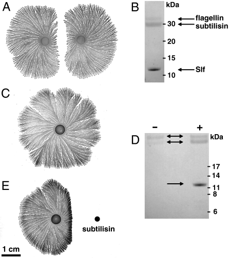
Fig. 1.
Colonies of P. dendritiformis (T morphotype) grown for one week on 1.5% agar with 2 g/L peptone, and analysis of proteins released into the medium. (A) Two sibling colonies grown after simultaneous inoculation of an agar plate at the same distance from the plate’s center. (B) Proteins extracted from the agar between two colonies, analyzed by SDS–PAGE gel electrophoresis and identified by peptide sequencing as flagellin at 32 kDa, subtilisin at 30 kDa, and Slf at 12 kDa (Arrows). (C) A single colony of P. dendritiformis grown on an agar plate. (D) SDS–PAGE electrophoresis of proteins extracted from agar surrounding a single colony, without (indicated by - or with +) added subtilisin, as shown in panels C and E. (E) A single colony with subtilisin (0.1 mg dissolved in 5 μL) added 1 d after inoculation (Black Dot).

