Fig. 2.
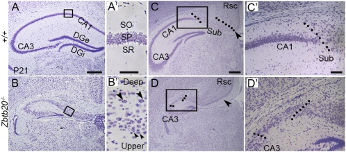
Abnormal cytoarchitecture of the Zbtb20−/− hippocampus. (A–D) Nissl-stained coronal forebrain sections at P21. (A′ and B′) High-magnification views of the boxed areas in A and B, respectively. The normally single layer of the stratum pyramidale (SP; A′) was transformed into two principal layers in the mutant CA1 field (B′): an upper layer (small arrowheads) and a deep layer (large arrowheads). (C and D) At a more caudal level, a subiculum-like structure appeared adjacent to the mutant CA3. Dotted lines indicate boundaries of subiculum. (C′ and D′) High-magnification views of the boxed areas in C and D, respectively. Sub, subiculum. (Scale bars: 400 μm for A–D; 50 μm for A′ and B′; 100 μm for C′ and D′.)

