Fig. 5.
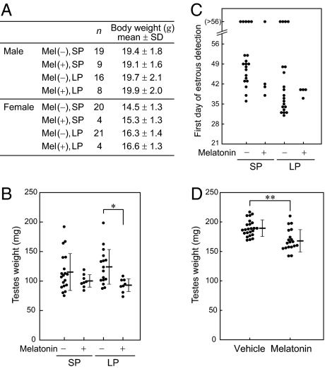
Effect of melatonin on mouse gonadal development. (A–C) The offspring of (B6J × MSM) × B6J were maintained in LD8:16 (SP) or LD16:8 (LP) after weaning. Melatonin productivity is estimated from the genotypes of Aanat and Hiomt. (A) Effect on body weight at 7 weeks of age. (B) Effect on male gonadal development. Testes were weighed at 8 weeks of age. Values from individual animals (circles) as well as mean ± SD values (horizontal bars) are shown. Testicular weights of melatonin-deficient males were significantly greater than those of melatonin-deficient mice (P < 0.01, ANOVA), and it was particularly prominent under LP (*P < 0.002, posthoc t test). (C) Effect on female gonadal development. The first day of predominant appearance of cornified epithelial cells in a vaginal smear is plotted. (D) The effect of exogenous melatonin on testis development of melatonin-deficient ICR mice. Testes were weighed at 5 weeks of age. Values from individual animals (circles) as well as mean ± SD values (horizontal bars) are shown. Testicular weights of vehicle-treated males were significantly greater than those of melatonin-treated males (**P < 0.001, t test).

