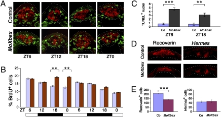
Fig. 3.
Xbsx knockdown leads to increased S-phase entry during the night and to a reduction of pineal photoreceptors. (A) Representative cryostat sections showing BrdU incorporation (green staining) in control- and MoXbsx-injected embryos during 24 h between stage 26 and stage 34. The Xotx5-positive area (red staining) is circled. (B) Quantification of BrdU-labeled cells present in the Xotx5-positive area of control-injected (blue bars) and MoXbsx-injected (red bars) embryos at eight time points. At each time point, the mean percentage per pineal organ of BrdU-positive nuclei over the total number of nuclei is plotted against ZT time. Data represent pooled results from three independent experiments. The number of cells counted is shown in Table S1. (C) Average number of TUNEL-positive nuclei in the Xotx5-positive area per pineal organ for embryos collected at ZT6 and ZT18, corresponding to stage 36 and stage 38, respectively. Data represent pooled results from three independent experiments. Six control (Co)- and six MoXbsx-injected embryos were analyzed per time point in each experiment. (D and E) Analysis of pineal cell types in control- and MoXbsx-injected stage 42 tadpoles. (D) Representative cryostat sections showing expression of markers for photoreceptors (Recoverin) and projection neurons (Hermes). (E) Average numbers of cells positive for Recoverin and Hermes per pineal organ. Data represent pooled results from three independent experiments. For each marker, four control (Co)-injected and four MoXbsx-injected embryos were analyzed per time point in each experiment. Asterisks indicate statistical differences as determined by the Student's t test: **P < 0.01; ***P < 0.001. Error bars indicate SEM.

