Fig. 4.
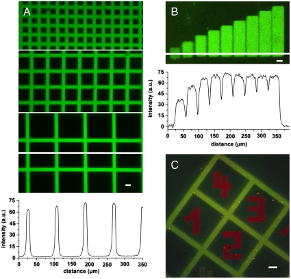
In situ assembly of proteins by light. (A) High contrast fluorescence images of Alexa488-MBP-H10 interacting with photo-patterned PA tris-NTA-His5 interfaces revealing sharp, diffraction limited edges. For photo patterning three distinct grid masks of different feature sizes were used. The white line indicates the position for which the fluorescence intensity profile is presented below the images. (B) Freely chosen regions of a PA tris-NTA-His5 interface were in situ exposed by laser scanning microscopy. Increasing time-dependent UV doses (exposure times 0–5 min) generate a protein gradient of Alexa488-MBP-H10 molecules. The corresponding fluorescence intensity profile emphasizes the stepwise increase in protein binding. (C) Orthogonal multiplexing of protein binding at a PA tris-NTA-His5 interface is demonstrated by using mask and in situ laser lithography in combination. The green pattern of Alexa488-MBP-H10 originates from the mask photo-patterned PA tris-NTA interface. Subsequently, in situ laser lithography was employed to write red numbers of ATTO565-MBP-H10 molecules into areas initially shadowed by the photo mask. The scale bars have a length of 20 μm.

