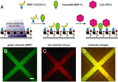
Fig. 5.
Two-dimensional organization of receptor-virus particle by light. (A) Schematic representation of capturing viral particles onto PA tris-NTA interfaces. The virus specific VLDL receptor (MBP-V33333-H6) doped with Alexa488-MBP-H6 was preimmobilized onto photo-patterned PA tris-NTA-His5 interfaces. Subsequently, Cy5-labeled HRV2 was bound by its receptor. Highly sensitive total-internal reflection fluorescence microscopy was used to detect the two-dimensional organization of receptor proteins (MBP-V33333-H6 was detected indirectly via cocapturing of fluorescent Alexa488-MBP-H6) (B) and Cy5-labled HRV2 particles (C). An overlay image of (B) and (C) is presented in (D). The scale bar has a length of 10 μm.

