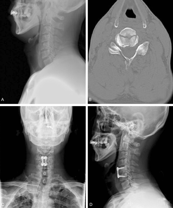
Fig. 2.
A 52 year old male patient with a C5 tear drop fracture (Case No.14). (A) The preoperative lateral roentgenogram shows anterior displacement of a bony fragment. (B) The CT shows a T-shape pattern with a sagittal fracture. (C) The postoperative radiograph shows the consolidation of the grafted bone. (D) The postoperative radiograph shows the consolidation of the grafted bone.

