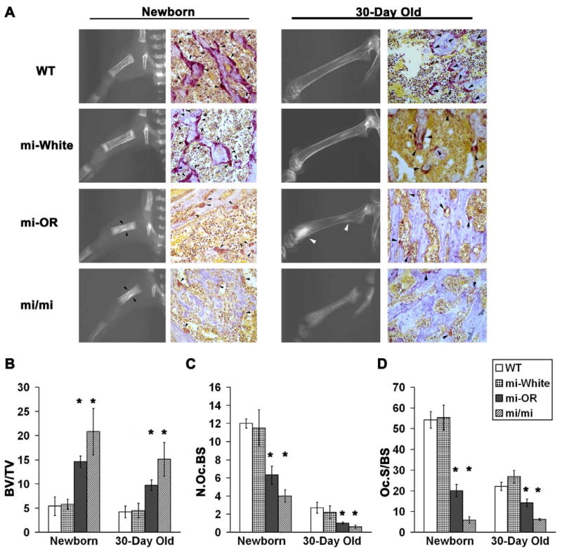
Figure 1. Mitfor/or mice exhibit osteopetrosis that improves with age.

A. Representative (see below) radiographic images of long bones (left in each panel) and digital microscopic images of Acp5 and hematoxylin stained 5 μM femur sections(right in each panel) from newborn (Left panel) and 30-day old (Right panel) WT, Mitfwh/wh (mi-White), Mitfor/or (mi-OR) and Mitfmi/mi (mi/mi) mice are shown. Black arrow heads in newborn Mitfor/or and Mitfmi/mi radiographs indicate sclerotic lesions in mid-diaphysis and white arrowheads in 30-day old Mitfor/or radiographs indicate sclerotic lesions in distal metaphysis of femurs. Black arrowheads in the Acp5 stained sections indicate osteoclasts positive for Acp5 enzyme activity. B-D. Bone and osteoclast parameters measured by histomorphometric analysis of Acp5 bone sections from new born and 30-day old WT, Mitfwh/wh (mi-White), Mitfor/or (mi-OR) and Mitfmi/mi (mi/mi) mice are shown. B: BV/TV indicates the percentage of unresorbed trabecular bone volume to total bone surface; C:- N.OcS/BS indicates the number of osteoclasts per total bone surface and D:- Oc.S/BS indicates the percentage of osteoclast surface to total bone surface. For all three analyses, averages of n≥30 for newborn mice and n=20 for 30-day old mice in the respective genotypes is shown. * indicates statistical significance with p < 0.05, Student's t-test.
