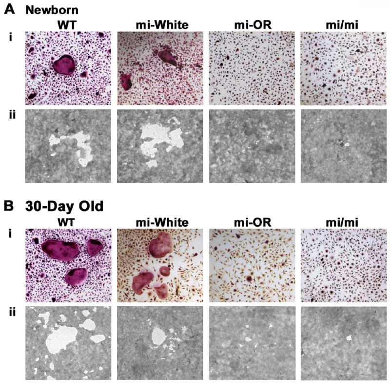
Figure 2. Mitfor/or precursors form fewer functional multinuclear osteoclasts than WT in vitro.

Precursors from either newborn (A) or 30-day old mice (B) of the indicated genotypes were cultured in the presence of CSF-1 and RANKL on gelatin coated wells for the formation of multinuclear osteoclasts (Ai and Bi) or on calcium phosphate coated wells for the formation of resorption pits (Aii and Bii). Representative digital microscopic images of Acp5 stained osteoclasts derived from newborn (Ai) or 30-day old (Bi) mice of the indicated genotypes. Figures Aii and Bii show representative digital microscopic images of resorption pits made by osteoclasts, from indicated genotype and age. Quantitation of the differentiation and functional assays is summarized in tables 1 and 2.
