
Figure 1.
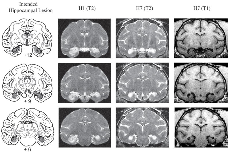
Left column shows coronal sections from a standard rhesus monkey brain depicting the intended hippocampal lesion (shaded region). Right columns show postoperative MR images for cases H1 and H7 at matching levels. T2-weighted MR images reveal the extent of white hypersignal, which reflects edema due to injections of excitotoxin and therefore the approximate site of the hippocampal lesions. T1-weighted MR images for monkey H7 shows gray matter – white matter contrast; note the marked shrinkage of the hippocampal formation bilaterally. Numerals indicate distance from interaural plane (0).