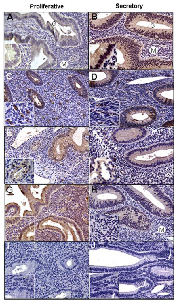
Figure 4.
Representative immunocytochemical localization (brown precipitate) of angiopoietin-1 (Ang-1; A, B), Ang-2 (C, D), Tie-2 receptor (E, F), and thrombospondin-1 (G, H) in the endometrium during the early (A, G) and late (C, E) proliferative and mid-late secretory (B,D,F,H) phases of the baboon menstrual cycle; (I) replacement of primary antibody with goat immunoglobulin G; (J) replacement of secondary anti-goat immunoglobulin in the Tie-2 reaction with secondary anti-mouse immunoglobulin. G, gland; M, microvessel. Final approximate magnification 200× in each panel, 640× in the inserts of panels A and B, and 400× in the remaining inserts.

