Figure 4.
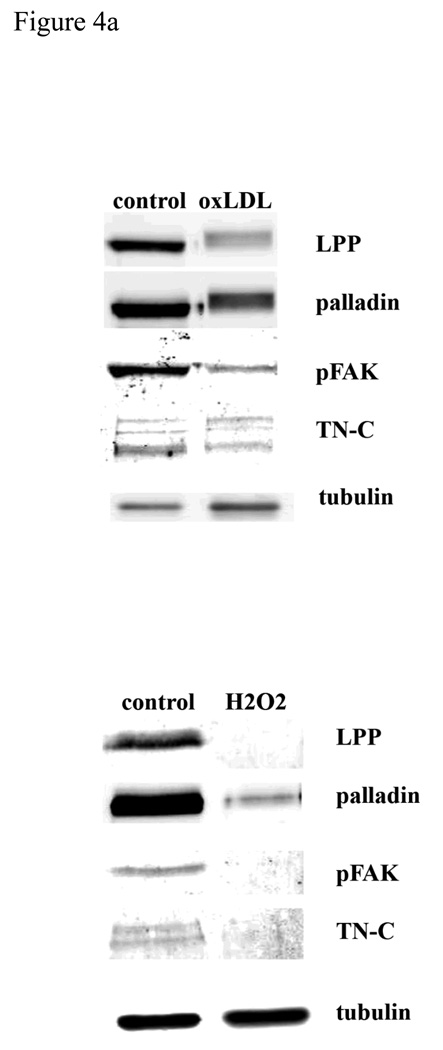
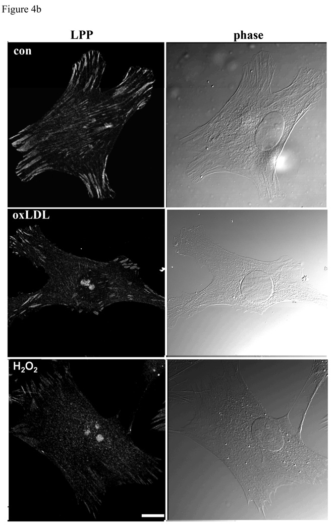
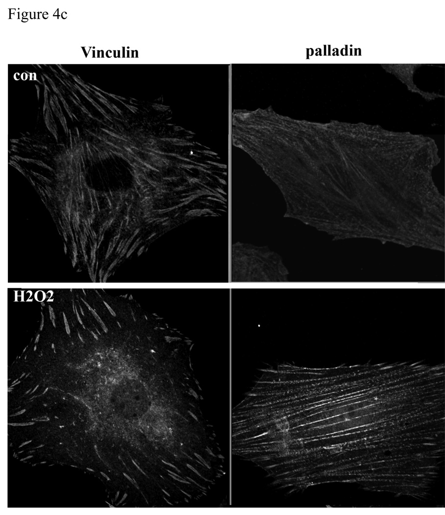
LPP, palladin, phosphoFAK and TN-C expression are down regulated by oxidative stress. A): R518 cells were treated with either 100ug/ml of oxLDL for 6h or 100µM of H2O2 for 24h, and cells were trypsinized and lysed for Western blotting. LPP, palladin, phosphoFAK and TN-C protein expression was significantly decreased from controls, p<0.05, n=3. Equivalent numbers of cells were loaded in each lane. Tubulin was used as loading control. B): LPP translocates to the nucleus, but not vinculin, in response to oxidative stress. Cells were treated with either 100ug/ml of oxLDL or 100µM of H2O2 for 1h, and cells were fixed for immunofluorescence staining with LPP antibody. Phase contrast image shows the cell morphology. Cells shown are representative cells of the population. Scare bar 50µm.


