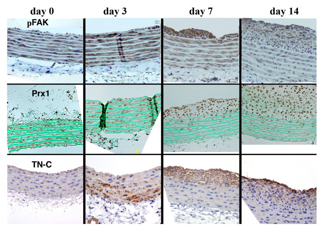
Figure 7.
Immunohistochemistry showing pFAK, TN-C and Prx1 expression in a time and spatial dependent manner in SMCs of the neointima and media of injured rat aortae at day 0 through to day 14. The vessel lumen is oriented to the top of each panel. For each experiment, there were 4 animals and 4 sections were observed. Similar patterns of immunolabeling were observed in each experiment for a given time point.

