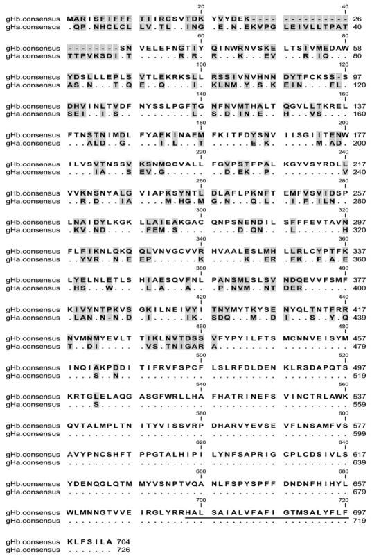
Figure 2.
Comparison of consensus amino acid sequences of gHa and gHb of RRV. Identical residues are indicated with dots. Gaps in the sequence are indicated by dashes. Shading indicates variable positions. The sequences were aligned using the Mafft program with default parameters. The putative membrane-spanning domain is underlined.

