
Figure 4.
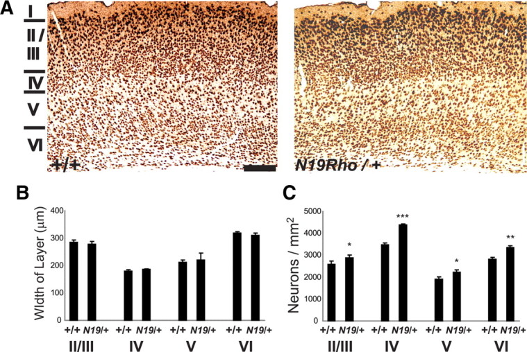
N19–RhoA mice show an increased density of neurons in somatosensory cortex. A, Immunohistochemical staining of somatosensory cortex in P65 wild-type (+/+) and N19–RhoA/+ mice, using an antibody recognizing NeuN. For each coronal section, dorsal is to the top, and lateral is to the right. Layers I–VI are indicated to the left. B, Quantification of the dorsoventral width of layers II/III, IV, V, and VI. C, Quantification of NeuN-positive neurons in layers II/III, IV, V, and VI. *p < 0.5, **p < 0.01, ***p < 0.001, Student's t test (n = 4). Scale bar: A, 200 μm.