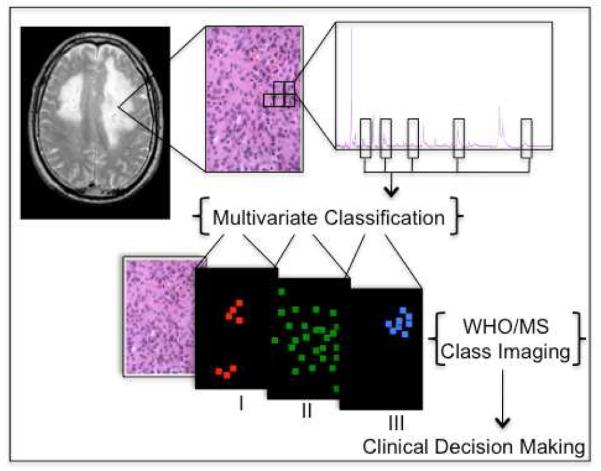
Scheme 1.

After a tissue biopsy is sectioned and stained, individual mass spectra are obtained uniformly across a sister tissue section surface. Spectra from tissues of different WHO grades are used to perform multivariate classification based on expert histopathology diagnosis. Classification models are then used to grade individual spectra from a tumor specimen, and the molecular images are rendered as class images. From these images, molecular indication of progression could be detected while still not observable by microscopic evaluation, and potentially contribute to clinical decision making.
