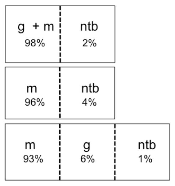
Figure 3.

General classification of meningioma images using a support vector machine trained on graded profiles. Combined with a non-tumor training set, several tumor sets were examined: meningiomas combined with gliomas, only meningiomas, meningiomas and gliomas separately. The ability to generally distinguish tumor from non-tumor under each scenario indicates the potential of this approach in a clinical setting. Labels indicate non-tumor brain (ntb), meningioma (m), and glioma (g), and percentages represent the distribution of total meningioma image spectra recognized by a given class.
