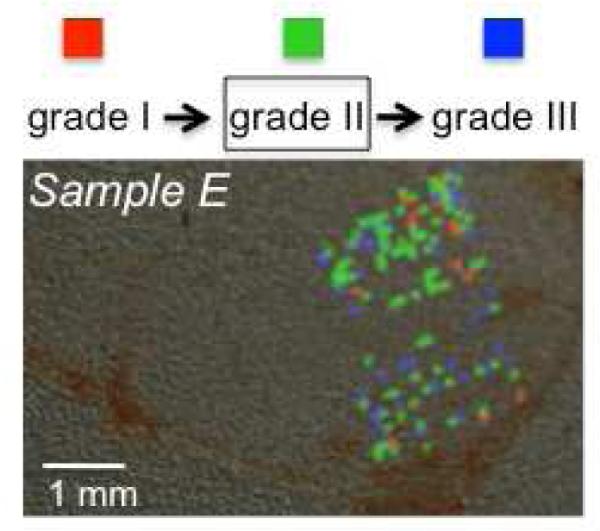
Figure 5.

Class imaging of a tissue specimen of histological grade II from a patient with progressive recurrence from grade I to II and to III shows the presence of regions of distinct grades. While many image spectra were discarded as null or noisy, the admissible spectra suggest spatial clustering of the different grade signals. Colored pixels represent Grade I (red), Grade II (green), Grade III (blue) regions.
