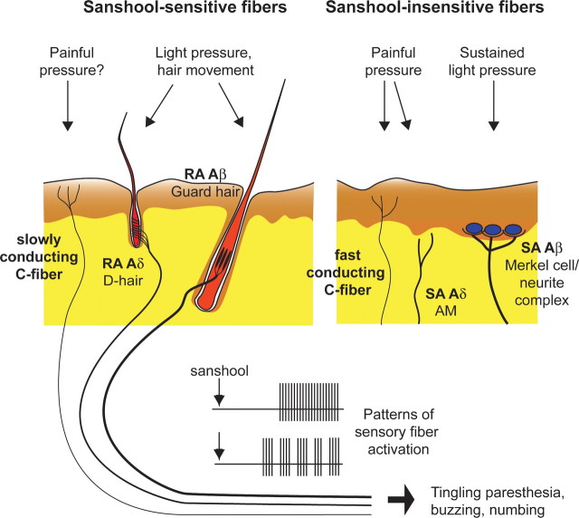
Figure 6.
Schematic of cutaneous mechanosensory afferents underlying sanshool-induced tingling paresthesia. The putative cutaneous innervation patterns of sanshool-sensitive (left) and sanshool-insensitive (right) mechanosensory afferents were examined in this study (Brown and Iggo, 1967; Burgess et al., 1968; Luo et al., 2009). The dermal and epidermal layers of the skin are represented in yellow and brown, respectively. Thickness of sensory fibers represents degree of myelination. Application of sanshool to the skin induces tonic or bursting activation of fibers, leading to sensation of tingling paresthesia (bottom).

