
Figure 2.
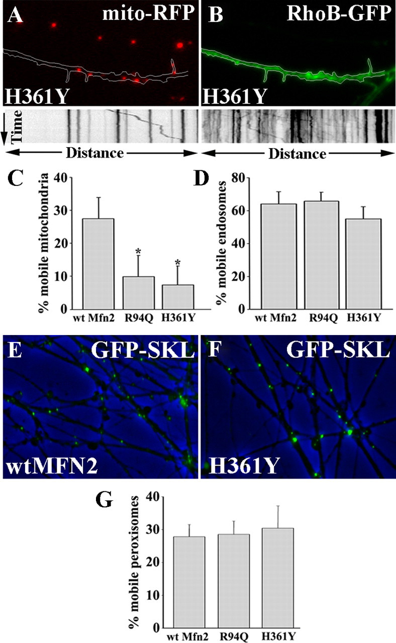
CMT2A-associated MFN2 mutants specifically disrupt mitochondrial transport. A, B, DRG neurons expressing the H361Y or R94Q MFN2 disease mutants were coinfected with mito-RFP (A) and RhoB-GFP (B) mitochondrial and endosomal markers, respectively. Mutant-expressing neurons revealed diminished mitochondrial mobility (A, kymograph) in the same axons that showed normal endosomal transport (B, kymograph). C, D, The percentage of mobile mitochondria is significantly decreased in R94Q- and H361Y- compared with wtMFN2-expressing neurons (*p < 0.001), whereas the percentage of mobile endosomes in these axons was normal. E, F, Overlay images of phase contrast and EGFP-SKL (which labels peroxisomes) in wtMFN2 (E)- and H361Y (F)-expressing DRG axons. G, Similar to endosomes, the percentage of mobile peroxisomes was unaltered by the expression of MFN2 mutants.