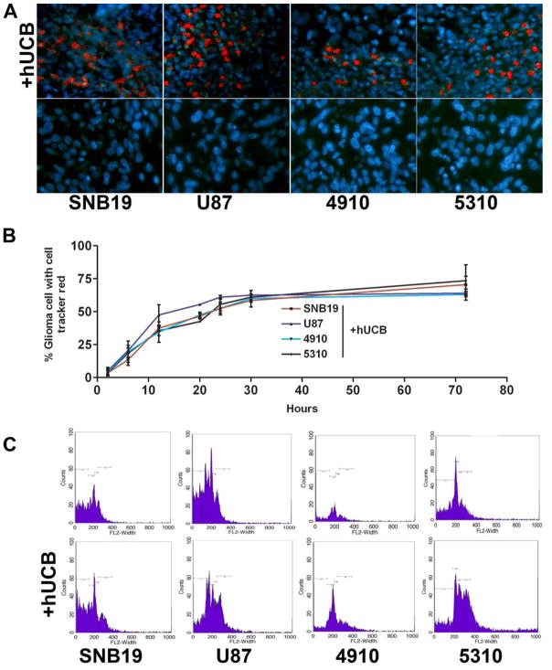
Figure 3.
To determine the interaction of hUCB with glioma cells, a modified Boyden’s chamber was used. The chamber consisted of a porous membrane (0.22 μm pore size), which was coated with Matrigel to facilitate the growth of cells on both the upper and lower surfaces. Human umbilical cord blood stem cells were grown on the upper surface and glioma cells (SNB19, U87, 5310 or 4910) were grown on the lower surface as previously described. The experiment was performed in multiple sets to determine cell-to-cell interaction over time. The cells were allowed to grow for 72 hrs under standard cell culture conditions. Periodic images were taken at different time intervals (6, 12, 20, 24 36 and 72 hrs) of the lower glioma cell surface after the hUCB layer was scraped off (A) and the percentage of glioma cells with cell tracker red stain were determined and plotted over time (B). After 72 hrs, glioma cells were trypisinized from the lower surface of the membrane and sorted for cell tracker red fluorescence by FACS analysis (C).

