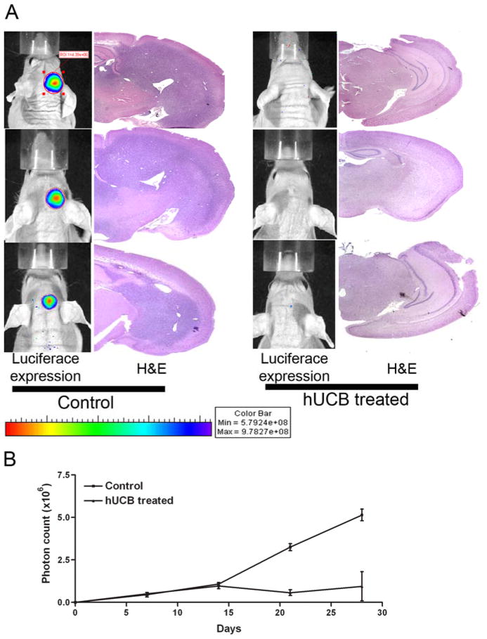
Figure 6.
To determine the anti-tumor effect of hUCB stem cells in in vivo conditions, we implanted nude mice with 1×10⁶ luciferase-expressing U87 cells. Seven days after implantation, mice were implanted again with 1×10⁵ hUCB cells or 10 μL of PBS at the site of U87 implantation. Tumor growth was monitored by periodic imaging with the Xenogen imaging system after intraperitoneal injections of luciferin solution as per standard protocols. Four weeks after treatment, mice were sacrificed and brains harvested followed by paraffin embedding and sectioning. The paraffin sections were H&E stained for visualization of tumor cells (A). Tumor burden was quantified as photon count and is graphically represented (B).

