
Figure 2.
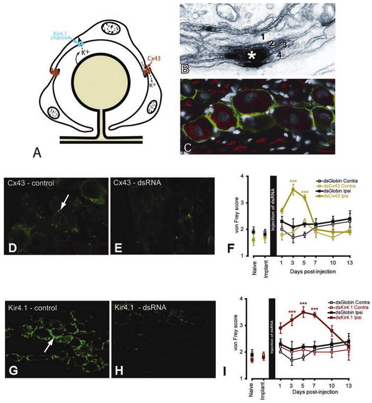
A, Diagram illustrating the role played by connexin 43 (Cx43) in intercellular transport and Kir 4.1 in the uptake of K+ ions. B, Cx43 postembedding electron micrograph staining (asterisk) showing the location of a gap junction between two satellite ganglion cells (SGCs). Where SGCs join there is commonly an interdigitation of processes from the SGCs (here labeled 1, 2, 3, 4). C, In the trigeminal ganglion, SGCs express the glia-specific K+ channel, Kir4.1 (green). Neurons (N) and axons are labeled with NF160 (red). D–I, Immunohistological and behavioral results of using dsRNA against Cx43 (D, E) and Kir4.1 (G–I). For each dsRNA, the control image (D, G) shows staining from an uninjected trigeminal ganglion and the second image (E, H) shows staining from a trigeminal ganglion 5 days after dsRNA injection. von Frey scores were measured for 13 days following injection of the dsRNAs (F, I). In each case there was a transient and reversible increase in von Frey scores for the active dsRNA but not for the control (globin) dsRNA nor on the side contralateral to the injection. *** P < .001, compared with contralateral side. (Additional information on protocols, statistical analysis, and results can be found in Ohara and others 2008; Vit and others 2008)