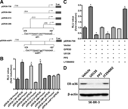
Figure 2.
GPR30 signaling activates ER-α36 promoter activity. A, Schematic structures of luciferase reporter plasmid driven by different 5′-truncated promoters of ER-α36. The −736, −584, −513, and −296 indicate residues upstream of the transcription initiation site, respectively. An AP-1-binding site, which was mutated in pER36-mAP-1, is also indicated. B, HEK293 cells were transfected with different reporter plasmids together with an empty expression vector or the expression vector for HA-GPR30, and luciferase activities were assayed and normalized using a cytomegalovirus-driven Renilla luciferase plasmid. Columns, Means of four independent experiments; bars, se. *, P < 0.05, for cells transfected with GPR30 vs. without GPR30. C, HEK293 cells were transfected with the pER36–736 reporter plasmid with the empty expression vector or the expression vector for GPR30 and then treated with vehicle, 10 μm U0126, 10 μm PP2, or 10 μm LY294002 for 24 h after which luciferase activities were analyzed. Results shown in the graph were mean ± se, and experiments were repeated four times. *, P < 0.05 for cells cotransfected with an empty expression vector and treated with vehicle vs. different conditions. D, SK-BR-3 cells maintained in DMEM supplemented with 10% FBS were treated with vehicle, 10 μm U0126, 10 μm PP2, or 10 μm LY294002 for 24 h. Cells were then harvested and analyzed by Western blot analysis. RLU, Relative light units.

