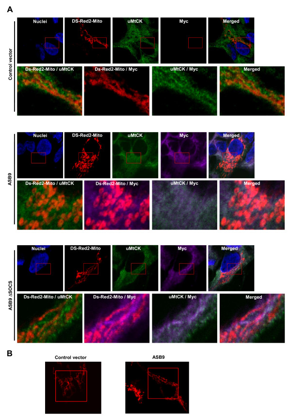
Figure 12.
Colocalisation of human ankyrin repeat and suppressor of cytokine signalling (SOCS) box protein 9 (ASB9) and ubiquitous mitochondrial creatine kinase (uMtCK) in mitochondria. The pDSRed2-Mito vector was transfected into human embryonic kidney 293 (HEK293) cells that stably express null (empty vector), Myc-tagged ASB9 or ASB9ΔSOCS. After 24 h incubation, the cells were fixed with 4% paraformaldehyde. (a) A confocal laser scanning microscopy was used to visualise the location of individual proteins with the aid of the mouse anti-Myc antibody (for ASB9, purple) and the goat anti-uMtCK antibody (green), respectively. DSRed2-Mito was expressed in mitochondria as a red colour. Cells were stained with Hoechst no. 33258 to visualise the nuclei (blue colour). (b) A confocal laser scanning microscope was used to visualise the structure of mitochondria with the aid of DSRed2-Mito. Three-dimensional movies of the figures are available as Additional files 1 and 2.

