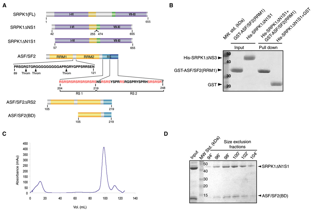
Figure 1. Isolation of the Core of the SRPK1:ASF/SF2 Complex.
(A) Domain organization of SRPK1 and ASF/SF2 and the fragments used for the complex formation and crystallization. I–XI denotes the substructures in the two kinase lobes as defined by Hanks and Quinn (1991). The putative thrombin cleavage sites in ASF/SF2 are shown by arrows.
(B) RRM1 does not interact with SRPK1. GST pull-down assay shows no interaction between SRPK1ΔN1S1 and RRM1 of ASF/SF2.
(C) Gel filtration elution profile of the complex. Size-exclusion chromatography shows that the purified complex of SRPK1ΔN1S1 and ASF/SF2(BD) is homogeneous in solution.
(D) SDS-PAGE analysis of peak fractions from size-exclusion chromatography.

