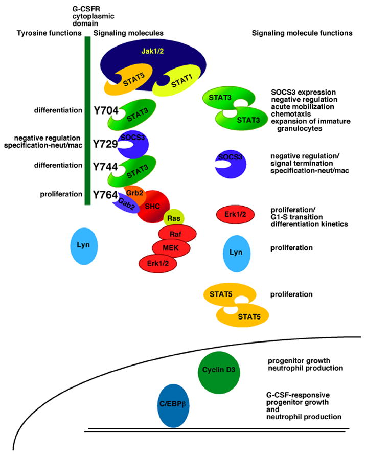
Figure 2.

Functions of G-CSFR tyrosine residues and receptor-activated signaling pathways. The intracellular region of the G-CSFR is illustrated (narrow green box, left; not drawn to scale), with the four tyrosine residues highlighted in black. On the left of each tyrosine residue are functions that have been assigned by in vitro and in vivo studies, including proliferation, differentiation and macrophage/granulocyte lineage-specification. On the right of each tyrosine, representations of signaling proteins that couple to specific residues are shown. The function of these molecules is listed on the right side of the figure. At the lower portion of the figure, a schematic diagram of the nucleus of a granulocytic progenitor is shown, along with additional signaling molecules that are required for ‘emergency’ granulopoiesis. References for tyrosine and signal protein function can be found in the text.
