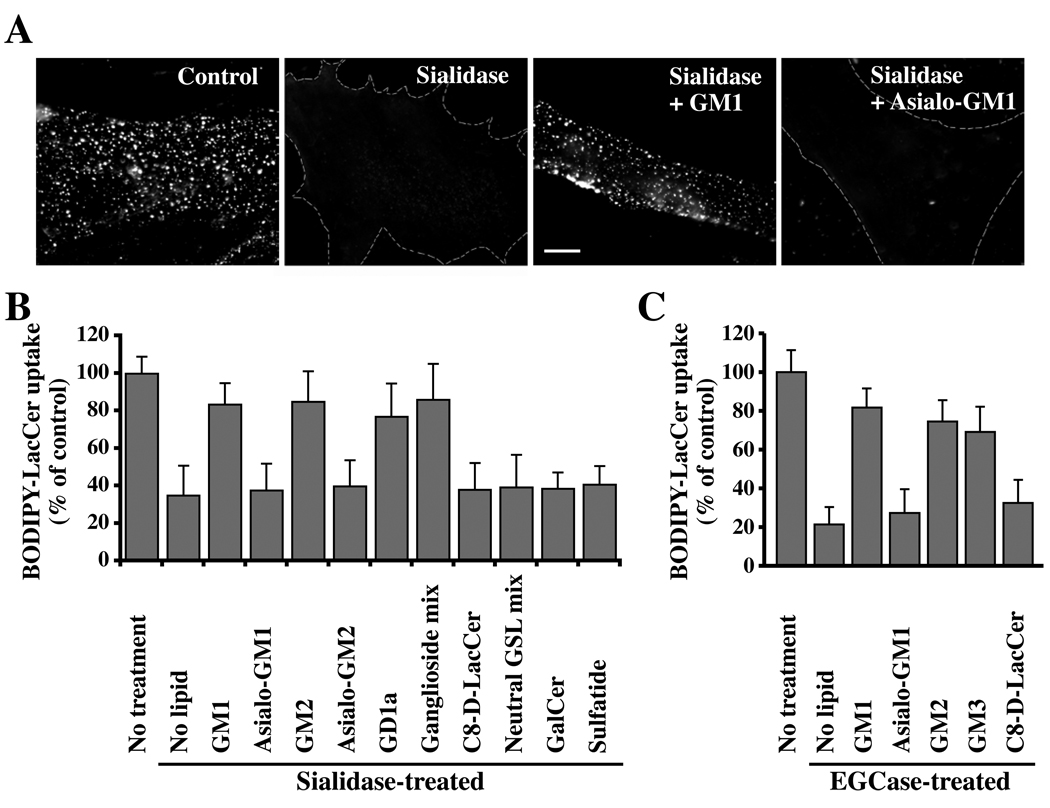
Fig. 2. Restoration of Bodipy-LacCer endocytosis in HSFs by sialic acid containing gangliosides.
Cells were pretreated with sialidase (A,B) or EGCase (C) as in Fig. 1, washed, and pulse-labeled with Bodipy-LacCer to monitor uptake after 5 min of endocytosis at 37°C. In some instances the enzyme-pretreated cells were incubated for 30 min at 10°C with the indicated non-fluorescent lipid, washed, and subsequently pulse-labeled with Bodipy-LacCer as above. (A) Fluorescence micrographs showing inhibition of Bodipy-LacCer uptake by sialidase and restoration of endocytosis by GM1 (but not asialo-GM1) ganglioside. Bar, 10 µm. (B,C) Quantitative results for restoration of Bodipy-LacCer endocytosis by various lipids after sialidase or EGCase treatments. “No treatment,” refers to no enzymatic pretreatment; “No lipid” shows effect of enzymatic treatment without lipid addition. Values were quantified as in Fig. 1 (n ≥ 30 cells/marker in 3 independent experiments) and are expressed as means ± SE relative to untreated control samples.

