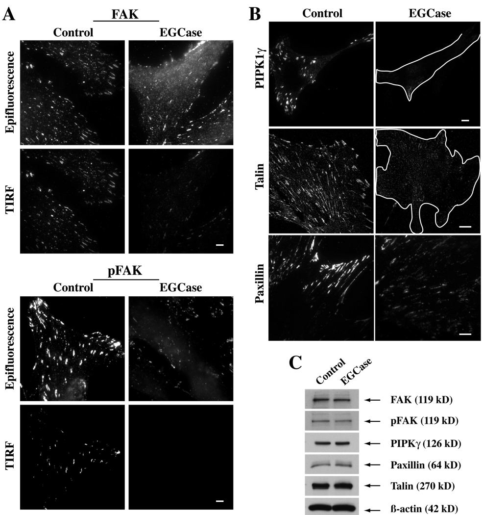
Fig 8. Effect of EGCase treatment of HSFs on focal adhesion components.
(A) Cells were grown in serum-containing medium, washed, and subsequently were untreated (Control) or pretreated with EGCase for 30 min at 37°C. The basal level of FAK and pFAK (Y397) was then assessed by antibody staining using both epifluorescence and TIRF microscopy. (B) Parallel cell samples, grown under the same conditions, were immunostained for talin or paxilin. In the case of PIPK1γ cells were transfected overnight with HA-tagged PIPKIγ prior to EGCase treatment, fixation and immunostaining. PIPKIγ and talin images were by confocal microscopy; paxilin images were by TIRF microscopy. (C) In parallel dishes, cells were untreated (Control) or pretreated with EGCase for 30 min at 37°C, lysed, and cell lysates (10 µg protein per lane) were immunoblotted for various proteins as indicated. Bars, 10 µm.

