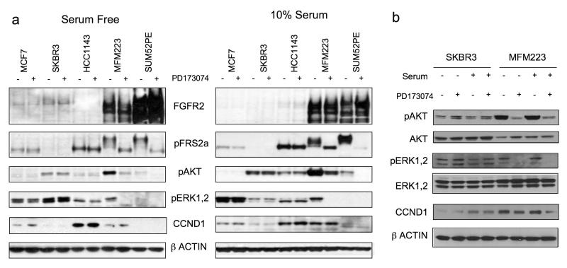
Figure 4.
a) Signalling downstream of FGFR2 in amplified cell lines. Indicated cell lines were grown either in 10% serum, or serum starved for 24 hrs, and lysates were made after 1hr exposure to 1μM PD173074 (+), or no exposure (−), as indicated. Lysates were subject to SDS-PAGE and western blotting with antibodies against FGFR2, phosphorylated FRS2-Tyr196, phosphorylated AKT1-Ser473, phosphorylated ERK1/2-Thr202/Tyr204, CCND1, and β-Actin. b) Side-by-side comparison of lysates from MFM223 grown in 10% serum or serum starved, with or without 1μM PD173074, with SKBR3 lysates for comparison.

