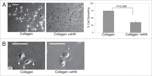
Figure 4.
Cell morphology changes dependent on HA in collagen I gels. (A) MDA-MB-231 cells (5 × 10⁴) were placed onto collagen matrix either immobilized with HMW HA or without HA in 20% FBS. Fourty-five minutes after placement of cells on matrix, cells were imaged and spreading was quantified. Cells were imaged at 60X magnification while all cells (>150) within a 10X field of view were enumerated. Statistics were calculated using an unpaired student’s t-test. The error bars represent s.d. from three separate experiments. (B) Time lapsed video microscopy was obtained from cells seeded as described in (A) over 1.5 hrs, revealing filopodia formation (1st panel, white arrowheads) when on collagen and lamellipodia formation (3rd panel, white arrow) when on collagen + eHA. Scale bar indicates 25 µm.

