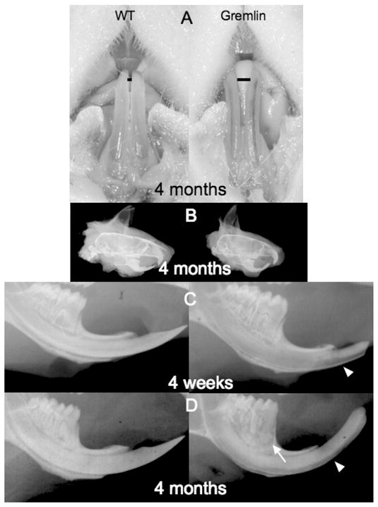
FIG. 1.

Gross appearance and radiographic analysis. (A) 4-month-old male Gremlin OE mouse (right panel) has pulp tissue visible through lower incisors. Also note widened space (black bar) between lower incisors in Gremlin versus. Wild type(WT) mouse. (B) Lateral cephalic view of 4-month-old male WT (left) and Gremlin (right) mice. Note the excessive curvature of upper and lower incisors of the Gremlin mouse. (C, D) Lower mandible radiographic examination, WT (left) Gremlin (right), 4 weeks (C), and 4 months (D). The pulp chamber in molars from 4 weeks Gremlin (C, right) showed significant enlargement compared with WT (C, left). Labial surface of Gremlin incisor is more radiolucent (C, D white arrowhead) and molars exhibit periapical alveolar bone resorption (D, white arrow).
