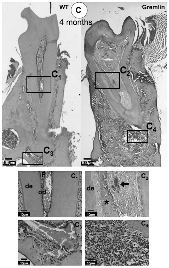
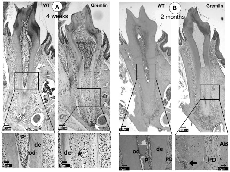
FIG. 2.

Histological analysis at 4 weeks (A), 2 months (B), and 4 months (C) of age. (A) At 4 weeks, ectopic calcification with entrapped cells is visible in the pulp space of the Gremlin mice (asterisk), which is seen as a tubular in nature in the enlarged view. Inflammation is seen at the Gremlin root apex (white arrow). The WT mouse pulp space is seen as normal. (B) Similar pulpal morphology is seen at 2 months, but with more extensive necrotic cells noted in root pulp (see enlarged view, arrow), with continued apical inflammation. Further, the PDL space of the Gremlin mouse appears to be much less cellular (see enlarged view Gremlin mouse). (C) As noted at earlier time points, 4 month Gremlin mouse pulp contains mineralized tissue with entrapped cells with extensive apical inflammation. C1–C4 Higher magnification of 4-month-old mice pulp and apical regions. C1 = Normal appearance of WT pulp and C2 = Entrapped cells in Gremlin mouse pulp (asterisk) with necrotic cells noted (black arrow). C3 = Periapical region of WT is normal in appearance. C4 = High degree of inflammation at the apex of Gremlin mainly composed of neutrophils, but lymphocytes and plasma cells are also present. Scale bars = 100 μm in whole molar images, 10 μm in enlarged images. De = dentin, od = odontoblasts, P = pulp, PD = periodontal ligament, AB = alveolar bone.

