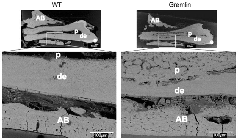
FIG. 3.

Backscatter mode scanning electron microscopy (SEM) image at age of 4 months old 1st molars in the left mandible. WT (left) mouse shows normal appearance of dentin (de), enamel (e), and pulp chamber (p). Gremlin mouse (right) molar exhibits thinner enamel with mineralized tissue in the pulp chamber. At higher magnification the mineral tissue appear bone-like, and also its presence results in diminished thickness of root dentin. Scale bars = 100 μm, AB = alveolar bone.
