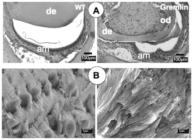
FIG. 4.

Histological and SEM analysis of 4 month lower incisors. (A) WT (left) has normal dentin thickness and labial enamel space with well-polarized ameloblasts observed. Gremlin OE mouse incisor (right) has much thinner dentin, and the enamel space is constricted mesiodistally with some ameloblasts lacking polarity. Scale bars = 100 μm, am = ameloblasts, de = dentin, od = odontoblasts. (B) SEM of WT enamel (left) shows characteristic weaved pattern of enamel crystal rods interpacked with interrod crystals. Gremlin enamel (right) lacks proper rod-interrod packing structure. Scale bars = 1 μm.
