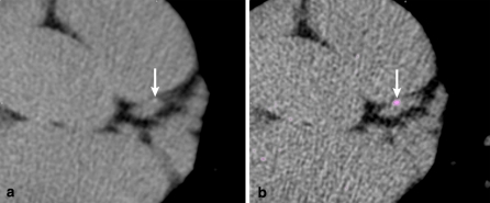
Fig. 2.
Calcium score in a 59-year old male with 3.0 mm (a) and 0.5 mm slice reconstructions. The Agatston score obtained at 3.0 mm reconstructions was zero as the visible calcium spot fell below the threshold value. With 0.5 mm slice reconstruction, the calcified lesion identified in the left anterior descending artery resulted in an Agatston score of 5

