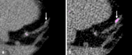
Fig. 3.
Calcium score of a 47-year old male with 3.0 mm (a) and 0.5 mm slice reconstructions (b). Identification of a calcified lesion in the left anterior descending artery at 0.5 mm reconstruction with an Agatston score of 9 (b, arrow), that fell below the threshold value for detection at the 3.0 mm reconstruction (a, arrow)

