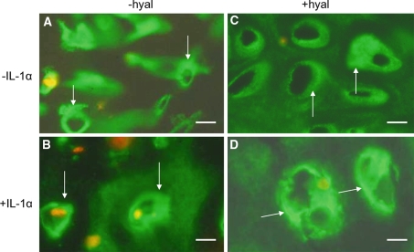
Fig. 7.
Localization of collagen type VI in sections from nasal cartilage explants cultured for 2 weeks with or without IL-1α. The sections were treated with or without hyaluronidase. a, b without hyaluronidase treatment (−hyal) c, d with hyaluronidase treatment (+hyal) a, c 2 week culture period without IL-1α (−IL-1α) b, d 2 week culture period with IL-1α (+IL-1α) Type VI collagen staining is present in a broad area around the chondrocytes (arrows). Treatment with hyaluronidase (c) resulted in a smaller but more intense staining area in between the chondrons (arrows). After a 2 week culture period with IL-1α (b, d), the distribution of type VI collagen is similar to that found in c following hyaluronidase treatment. Also here an intensively stained narrow area around the chondron (arrows) is next to a narrow unstained area. Interchondrally the staining becomes more intense after IL-1α incubation or hyaluronidase treatment. After 2 weeks culturing with IL-1α the explant is nearly completely digested. Bars: 0.2 μm

