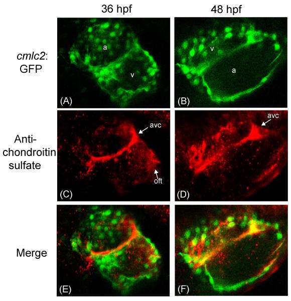
Figure 2.
Confocal images of Danio rerio hearts stained with an anti-chondroitin sulfate antibody at 36 and 48 hours post fertilization (hpf). (A,B) Confocal microscopy of Danio rerio containing the myocardium specific cmlc2:GFP marker (green). (C,D) Confocal microscopy of the same Danio rerio images stained with a CS-specific antibody (red). (E,F) Merged images. a= atrium; v= ventricle; avc = atrioventricular canal forming region; oft = outflow tract.

