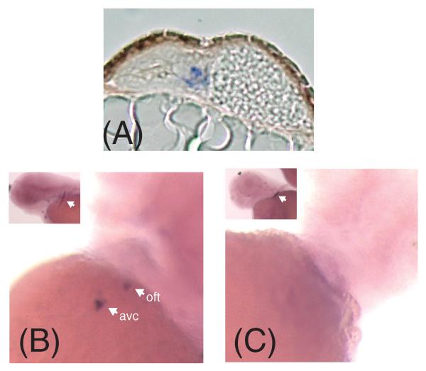
Figure 5.
Depletion of chondroitin sulfate leads to changes in the cell migration marker spp1. (A) In situ hybridization section demonstrating that the zebrafish osteopontin marker spp1 is localized to the atrioventricular canal endocardium in zebrafish. (B) spp1 is normally expressed in the atrioventricular canal (avc) and outflow tract (oft). (C) DX-treatment from 7 to 48 hpf results in the disappearance of spp1 in the AVC and OFT at 72 hpf. Note that while spp1 expression is absent in DX treated embryos, the presence of a stripe of spp1 expression at the base of the pectoral fin demonstrating the success of the in situ protocol (white arrow in inset of B and C).

