
Figure 2. Connections between primary afferent fibers and the spinal cord.
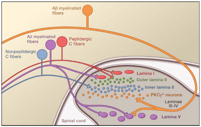
There is a very precise laminar organization of the dorsal horn of the spinal cord; subsets of primary afferent fibers target spinal neurons within discrete laminae. The unmyelinated, peptidergic C (red) and myelinated Aδ nociceptors (purple), terminate most superficially, synapsing upon large projection neurons (red) located in lamina I. The unmyelinated, non-peptidergic nociceptors (blue) target small interneurons (blue) in the inner part of lamina II. By contrast, innocuous input carried by myelinated Aβ fibers terminates on PKCγ (yellow) expressing interneurons in the ventral half of the inner lamina II. A second set of projection neurons within lamina V (purple) receive convergent input from Aδ and Aβ fibers.