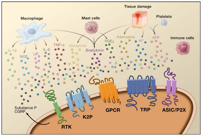
Figure 4. Peripheral mediators of inflammation.

Tissue damage leads to the release of inflammatory mediators by activated nociceptors or non-neural cells that reside within or infiltrate into the injured area, including mast cells, basophils, platelets, macrophages, neutrophils, endothelial cells, keratinocytes, and fibroblasts. This “inflammatory soup” of signaling molecules includes serotonin, histamine, glutamate, ATP, adenosine, substance P, calcitonin-gene related peptide (CGRP), bradykinin, eicosinoids prostaglandins, thromboxanes, leukotrienes, endocannabinoids, nerve growth factor (NGF), tumor necrosis factor α (TNF-α), interleukin 1β (IL-1β), extracellular proteases, and protons. These factors act directly on the nociceptor by binding to one or more cell surface receptors, including G protein coupled receptors (GPCR), TRP channels, Acid-sensitive ion channels (ASIC), two-pore potassium channels (K2P), and receptor tyrosine kinases (RTK), as depicted on the peripheral nociceptor terminal.
