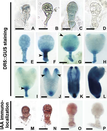
Fig. 2.
Localization of IAA in tobacco zygotes and proembryos. (A–C, E–L) Auxin-dependent DR5::GUS expression in zygote, 2-celled, 8-celled, early globular, middle globular, late globular, transition-stage, heart-shaped, early torpedo-shaped, late torpedo-shaped, and mature proembryos, respectively. (M–O) Immunoenzyme localization of IAA in the wild-type zygote, 2-celled, and early globular proembryos, respectively. (D, P) The control embryos in wide-type plants with GUS staining (D) or without the primary antibody (P), respectively. Arrowheads indicate the suspensor, cotyledon primordium regions, hypophysis cell region, and provascular strands in (F, G, I, J), respectively. GUS staining in blue (A–C, E–L). IAA signal in brown (M–O). (A–D, M–P) Bar=20 μm; (E–H) bar=40 μm; (I–L) bar=60 μm. (This figure is available in colour at JXB online.)

